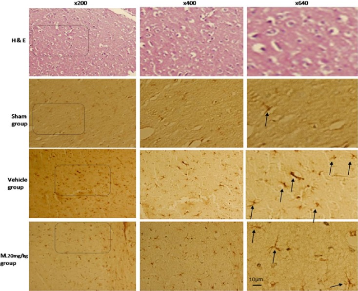
Figure 4.

Immunohistochemical analysis of glial fibrillary acidic protein (GFAP) positive cells in brain cortex of rat. Arrows show the GFAP positive cells. Bar=10 µm

Immunohistochemical analysis of glial fibrillary acidic protein (GFAP) positive cells in brain cortex of rat. Arrows show the GFAP positive cells. Bar=10 µm