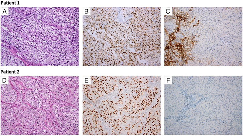
Figure 2.
H&E and Immunohistochemical staining with PAX8 and CA-IX. H&E staining demonstrating eosinophilic cells with voluminous cytoplasm from the subcarinal lymph node and anterior neck metastasis in patients 1 and 2, respectively (A and D). Positive nuclear PAX8 staining is present in both samples (B and E). CA-IX staining is absent in tumour cells (C and F), but present in the normal stromal cells of patient 1's lymph node sample (C).

