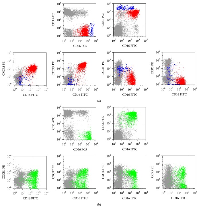
Figure 3.
Illustrative dot plots showing the expression of the CXCR1, CXCR2, CXCR3, and CCR5 chemokine receptors (CKR) in normal peripheral blood (PB) (a), where only CD56+low (red dots) and CD56+high (blue dots) are observed, and in the PB of a patient with a chronic lymphoproliferative disorder of NK-cells (CLPD-NK) (b), exhibiting a transitional CD56+int phenotype (green dots); other lymphocytes are shown in gray. In order to obtain the dot plots showed in this figure, the PB cells were stained with APC-conjugated anti-CD3, PC5-conjugated anti-CD56, PE-conjugated anti-CKR (CXCR1, CXCR2, CXCR3, or CCR5), and FITC-conjugated anti-CD16 monoclonal antibodies. As shown in (a), in the normal PB most CD56+low NK-cells are CXCR1+ and CXCR2+, whereas only a very small fraction of cells stains positively for CXCR3 and/or CCR5; in contrast, most CD56+high NK-cells are CXCR3+ whereas CCR5 is expressed in only a fraction and CXCR1 and CXCR2 are virtually negative. As it can be seen in (b), the expanded NK-cells from this patient, which have relatively high levels of CD56 expression, are CD16+ and they express the CXCR1, CXCR2, CXCR3, and CCR5 molecules in a considerable fraction of cells.

