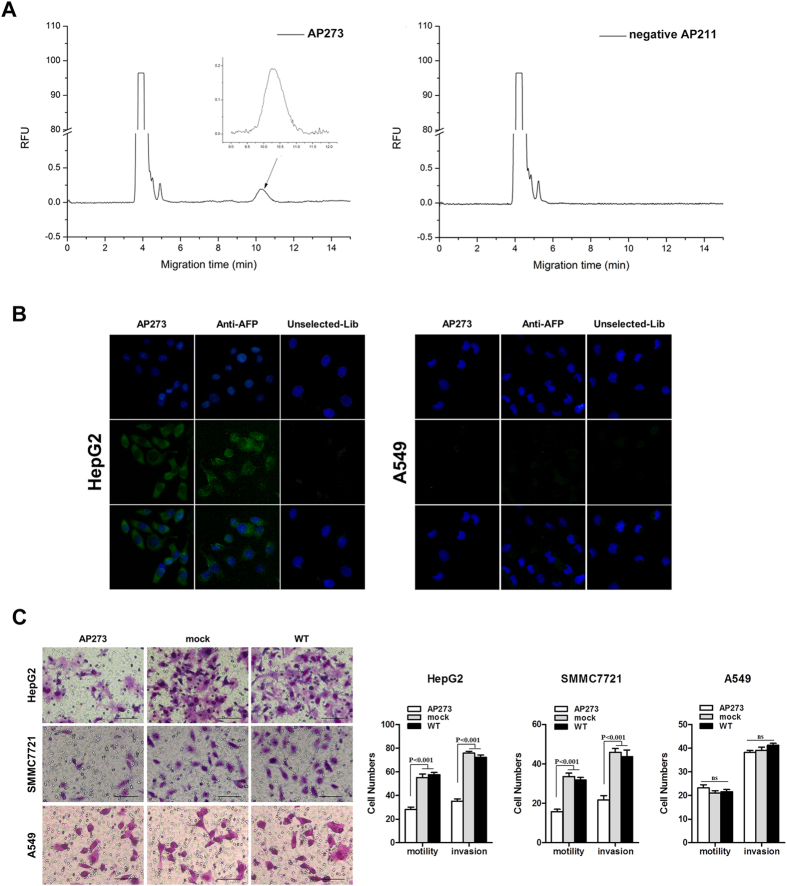
Figure 4. Binding specificity and functional evaluation of AP273.
(A) Capillary electropherograms of FAM-labeled AP273 and AP211. AP273 specifically bound AFP, while AP211 did not in CE assays. (B) Fluorescence imagings of HepG2 and A549 cells stained by FAM-labeled AP273, anti-AFP antibody and FAM-labeled unselected aptamer library, respectively. About 10 μg/ml of aptamer or antibody as the final concentration was used. DAPI was used for contrast staining (blue). (C) Representative images (left) and statistical results (right) of cell migration and invasion. HepG2, SMMC7721 and A549 cells were transfected with AP273, AP211 (mock) or nontransfected (WT), respectively. Bars, 100 μm. Data are mean ± SD and representative of three independent experiments.

