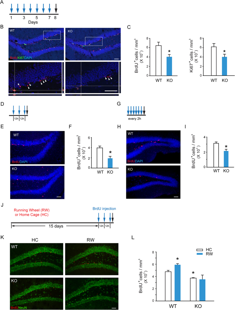
Figure 1. Decreased Proliferation of Neural Precursors in the SGZ of Adult β-arr1 KO mice.
β-arr1 KO mice (KO) and wild type littermates (WT) of 2–3 month old were used. (A–I) Mice were injected intraperitoneally with 100 mg/kg BrdU (blue arrows) and sacrificed (black arrows) at time points indicated (A,D,G). Sample projected confocal images were shown (B,E,H) and stereological quantification (C,F,I) of BrdU-positive and Ki67-positive (C) cells in SGZ of these mice were performed. Arrowheads indicate the localization of BrdU+ cells. Orthogonal views are samples of BrdU+ Ki67+ cells. The cell number was normalized to the GCL (granular cell layer) volume (in mm3). Data represent mean ± s.e.m.; n = 3 or 4 mice for each genotype; t- test, *p < 0.05. Scale bar, 50 μm; (J–L) After running wheel training (RW) or housed in home cage (HC) for 15 days, mice were injected with BrdU for 3 times (blue arrows) with an 12 h interval and sacrificed (black arrow) 2 h after the last BrdU injection (J). Immunostaining (K) and stereological quantification (L) of BrdU+ cells in SGZ were performed. The cell number was normalized to the GCL volume (in mm3). Data represent mean ± s.e.m.; n = 3 mice/group; *p < 0.05 vs. WT/HC, two-way ANOVA, post hoc Tukey test; Scale bar, 50 μm.

