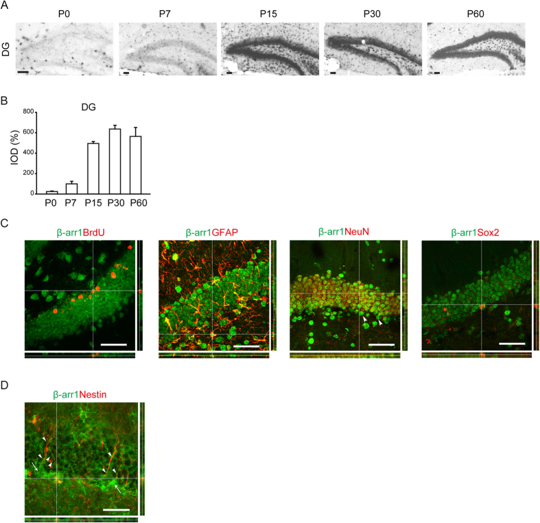
Figure 2. Expression of β-arr1 in Neural Precursors and Niche Cells of SGZ.
(A,B) Representative images (A) and data of optical densities (OD) (B) of in situ hybridization using β-arr1 antisense probe on brain sections from P0, P7, P15, P30, or P60 WT mice. Scale bar, 50 μm. (C) In situ hybridization with β-arr1 antisense probe combined with immunostaining for BrdU, GFAP, NeuN, and Sox2 in the DG on brain sections of 3-month-old WT mice. Orthogonal views are shown to confirm colocalization. Arrowheads indicate β-arr1+ NeuN− cells. (D) Co-immunostaining for β-arr1 and Nestin. Orthogonal views are shown to confirm colocalization. Arrows indicate the cell bodies of Nestin+ RGLs and arrowheads indicate their branches. Scale bar, 50 μm.

