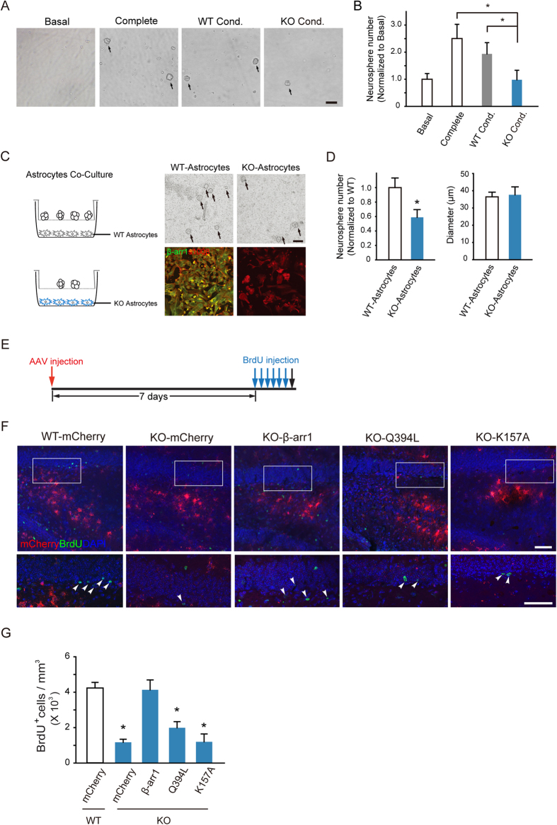
Figure 5. Nuclear β-arr1 Regulates the Proliferation of Adult Neural Precursors in DG.
(A,B) Tissue-conditioned media derived from DG of β-arr1 KO mice sustained less WT neurospheres than the complete media and the tissue-conditioned media derived from DG of WT mice. Data are normalized to basal value, n = 3 independent experiments. One-way ANOVA; Scale bar, 100 μm. (C,D) WT neurospheres co-cultured with β-arr1 KO DG astrocytes (KO-Astrocytes) were less than those co-cultured with WT (WT-Astrocytes). Data are normalized to WT, all error bars show s.e.m. of triplicated cultures (3–4 samples per group), t-test; Scale bar, 100 μm. (E–G) The schematic of experimental schedule (E), sample projected confocal images (F), and stereological quantification (G) of BrdU+ proliferating cells (arrowheads) in the SGZ of WT mice injected with AAV-hGFAP-mCherry (WT-mCherry) or β-arr1 KO mice injected with AAV-hGFAP-mCherry (KO-mCherry), AAV-hGFAP-β-arr1-mCherry (KO-β-arr1), AAV-hGFAP-β-arr1Q394L-mCherry (KO-Q394L) or AAV-hGFAP-β-arr1K157A-mCherry (KO-K157A). n = 3–6 for each group; Data represent mean ± s.e.m.; *p < 0.05 vs. WT-mCherry; one-way ANOVA, post hoc Tukey test; Scale bar, 50 μm.

