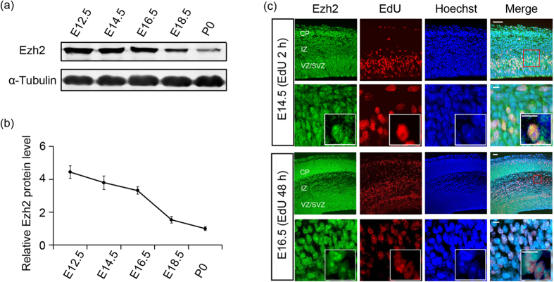
Figure 1. Ezh2 is expressed in both proliferating and postmitotic migrating neurons.
(a) Immunoblotting for Ezh2 in mouse cortex lysates at the indicated ages shows that Ezh2 is expressed in both early and late stages of cortical development (cropped images, and full-length blots are presented in Supplementary Fig. S3a online). (b) Quantification of Ezh2 expression level done by normalization to that of α-Tubulin. n = 3 for each stage. Data represent mean ± SEM. (c) Two hours after EdU administration, EdU+ proliferating cells are strongly immunostained with Ezh2 antibody. Two days after the administration, Ezh2 is continuously expressed by EdU+ migrating cells. Scale bars: 50 μm and 10 μm (insets).

