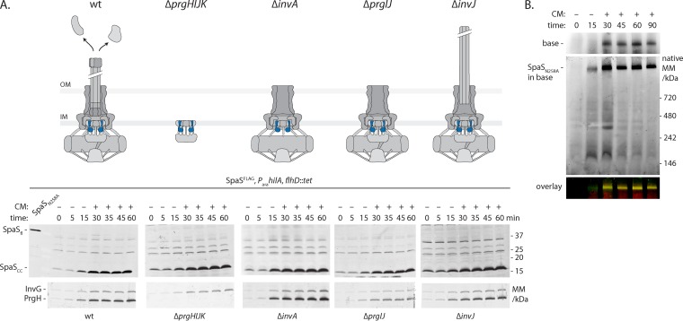
FIG 3 .
SpaS autocleavage is not regulated and occurs before needle complex assembly. All experiments shown were done with S. Typhimurium strains containing C-terminally FLAG-tagged SpaS (SpaSFLAG) and an arabinose-inducible SPI-1 master regulator HilA on the chromosome. (A) To assay the kinetics of SpaS autocleavage in wt and indicated mutant strains, bacteria were grown for 3 h, after which SPI-1 was induced by the addition of arabinose. Thirty minutes after induction, further protein synthesis was inhibited by the addition (+) of chloramphenicol (CM). Samples were taken at the indicated time points, and SpaS autocleavage was analyzed by SDS-PAGE, Western blotting, and immunodetection of its C-terminal FLAG tag. As a reference for the position of full-length SpaS, a sample of the autocleavage mutant was run in the leftmost lane. Induction of the needle complex base components InvG and PrgH is shown as a reference. The state of needle complex assembly and secretion in a given mutant is illustrated. The positions of molecular mass markers (in kilodaltons) are indicated to the right of the gel. SpaS is highlighted in blue. Abbreviations: OM, outer membrane; IM, inner membrane; SpaSfl, full-length SpaS. (B) To assay the kinetics of needle complex assembly, crude membranes of bacteria harvested at the indicated time points after SPI-1 induction were solubilized by DMNG, after which protein complexes were separated by blue native PAGE. Complex association of needle complex base components and SpaS was analyzed by Western blotting and immunodetection using an Li-Cor Odyssey system. Base components were detected in the 700-nm channel, while SpaSFLAG was detected in the 800-nm channel. The overlay of both channels is shown.

