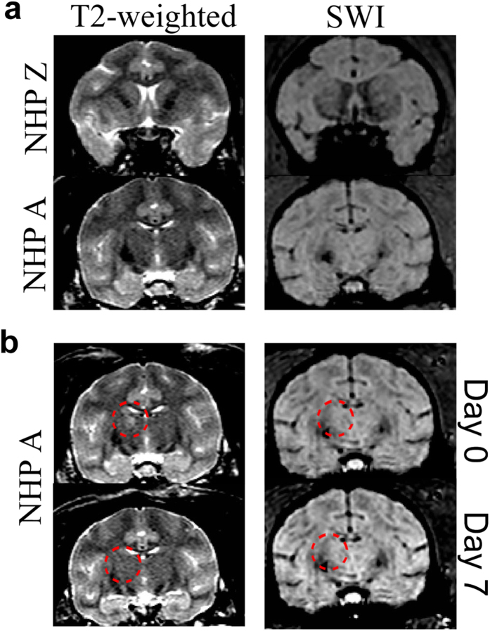
Figure 3. MRI Safety Verification.
T2-weighted and SWI sequences were acquired to verify the safety of the alert FUS procedures. (a) shows typical cases of both T2-weighted and SWI scans of the targeted regions. There were no abnormal hyper- or hypointense voxels present in any of the targeted regions (caudate, putamen or thalamus). (b) shows the one case for NHP A when there were hyperintense voxels in the targeted region in the T2-weighted scan, denoted by the red dashed circle. This area of hyperintense voxels could indicate the presence of edema. By day 7 the hyperintense voxels were absent. Neither on day 0 nor 7 were any hyper or hypointense voxels detected in the target regions of the SWI scans.

