
Figure 6. Cavitation Doses.
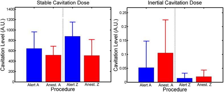
During both the alert and anesthetized FUS procedures, the passive cavitation detection recorded the signals emitted from the MB in the focal area of the transducer. Blue bars indicate average cavitation dosage and 95% confidence interval of the mean for alert FUS procedures while red indicates average cavitation dosage 95% confidence interval of the mean for anesthetized FUS procedures. (a) shows the average stable cavitation doses while (b) shows the average inertial cavitation dosages. For both NHP there was a non-significant increase in stable cavitation doses between the alert and the anesthetized FUS procedures. The inverse occurred with the inertial cavitation dose with smaller doses detected during the alert compared to the anesthetized FUS procedures.