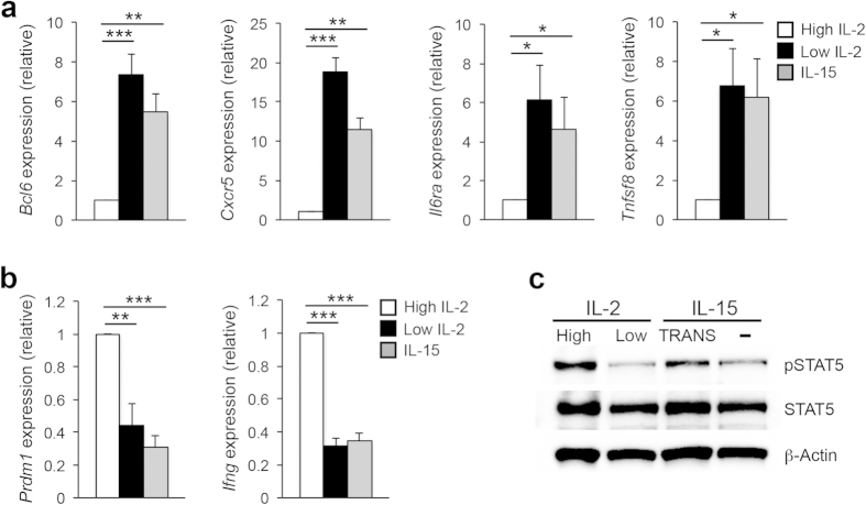
Figure 1. IL-15 treatment alone is insufficient to activate STAT5 and the TH1 program.
(a,b) Primary CD4+ T cells were isolated from C57BL/6 mice and stimulated on plate-bound αCD3 and αCD28 for 3 days under TH1-polarizing conditions. On day 3, cells were split and cultured with either high IL-2 (250 U/ml), low IL-2 (10 U/ml), or IL-15 (15 ng/ml). Two days following cytokine treatment, RNA was isolated to determine transcript levels for the indicated TFH and TH1-associated genes. Samples were normalized to Rps18 as a control. Data are presented relative to the high IL-2-treated sample. Three (a,b) independent experiments were performed with the error bars representing SEM. *P < 0.05, **P < 0.01, ***P < 0.001 (unpaired Student’s t-test). (c) Cells were treated and harvested as in “a” with the exception that an additional sample was treated with 15 ng/ml of trans-presented IL-15 (IL-15, TRANS). Following cell isolation, an immunoblot assay was performed to assess STAT5 activation levels. Total STAT5 and β-Actin are shown as controls. The image shown is representative of three independent experiments performed.

