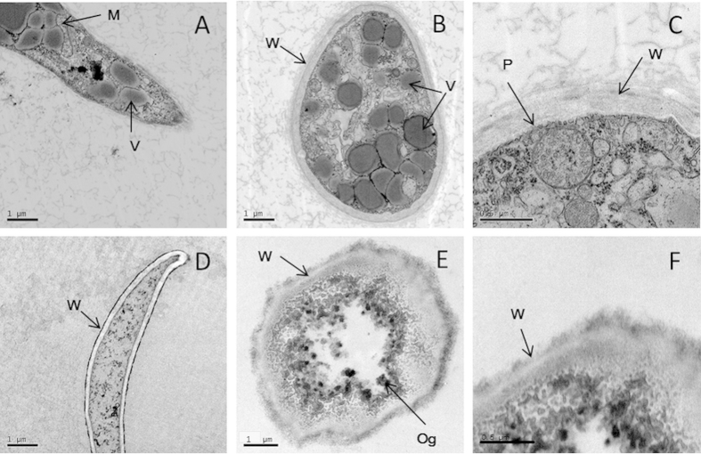
Figure 5. Transmission electron micrographs for the hyphae of P. litchii grown for 5 days on CAM plates with or without ZMS at 25 °C.
(A–C) control; (D–F) 2 μg/mL ZMS; (A,D) longitudinal section through the hyphae of P. litchii ( × 12,000); (B,E) tangential section through the hyphae ( × 12,000); (C,F) cell wall and plasma membrane of the hyphae ( × 20,000). M, mitochondria; P, plasma membrane; V, vacuoles; and W, cell wall.

