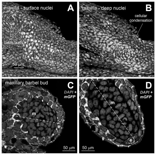
Figure 2. Ectodermal and mesodermal arrangements in the maxillary barbel bud.
All images are 3D renderings of confocal Z-stacks from fixed tissues. All cells are stained with DAPI (gray) to show nuclear morphology and cell arrangement. In all panels, anterior/proximal is to the left.
A) Lateral view of the surface of a developing maxilla from a juvenile zebrafish (standard length < 10 mm) at the prospective site of barbel outgrowth. Taste buds appear as cellular rosettes.
B) Deep confocal slice of the same specimen. The presumptive barbel is marked by a cellular condensation of dermal mesenchyme.
C) Optical section through an early maxillary barbel bud (~100 μm). The proximal-distal axis is dotted, with distal to the lower right. In this transgenic specimen, epithelial cells (e) express membrane-bound EGFP (white). The epithelial layer of the barbel bud (e) contains 2–3 layers of cuboidal or slightly flattened cells. The mesenchymal cells (m) form a whorl around the bud center.
D) Optical section through an elongating barbel bud (~300 μm). At this stage, the distalmost epithelial cells (e) are more squamous, with tangentially flattened nuclei. e = epithelial cells; m = mesenchymal cells

