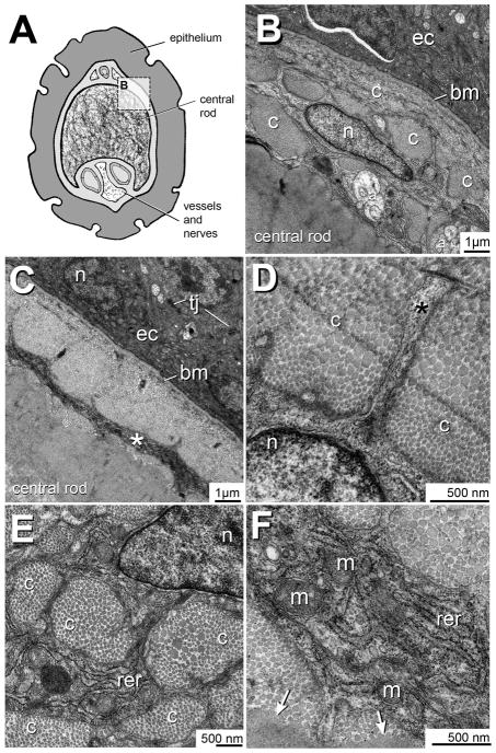
Figure 6. Transmission electron microcopy of the TCF+ ZMB core cells.
A) Cross-sectional overview of the adult zebrafish maxillary barbel. The rectangular boundary indicates the region magnified in panel B.
B) The maxillary barbel epidermal-dermal boundary. Ectodermal cells (ec) are electron-dense and rest on a prominent basement membrane (bm). Approximately 2 microns below this membrane lies the radially-flattened, pancake-shaped nucleus (n) of a TCF+ core cell. Adjacent to the cell are several collagen bundles (c). Deep to the cell are several axons (a) and the acellular matrix of the barbel’s central rod.
C) Micrograph of a similar region. The electron-dense epithelial cells (ec) are connected by tight junctions (tj). Approximately 2–3 microns below the basement membrane is a layer of cell cytoplasm (*) that appears as a continuous, circumferential ring.
D) A cytoplasmic projection (asterisk) extends from a matrix-bound cell through a dense field of collagen fibers (c).
E) Several cross-cut collagen bundles (c) near a TCF+ cell. The heterochromatic cell nucleus (n) is at the upper right. Between the bundles are ribbons of cytoplasm enriched in rough endoplasmic reticulum (rer). The small, dark nodules are individual ribosomes.
F) Magnification of a cytoplasmic ribbon shows extensive rough endoplasmic reticulum (rer) and intracellular vesicles. The double-wrapped ovoid organelles are mitochondria (m). Collagen fibers close to the cell appear well separated, showing individual ovoid cross-sections. In contrast, the collagenous matrix farther away from the cell appears hyperpolymerized (arrows), with no individual fibrils present.
a = axon; bm = basement membrane; c = collagen bundle; ec = ectodermal cell; m = mitochondria; n = nucleus; rer = rough endoplasmic reticulum; tj = tight junction.

