Figure 1.
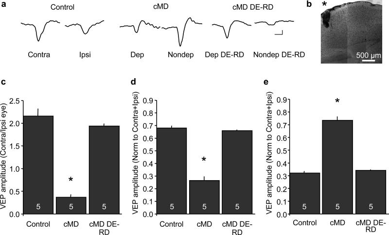
Regulation of layer IV VEP contralateral bias by chronic monocular deprivation. A. Representative VEP waveforms recorded from layer IV evoked by horizontal gratings (average of 100 presentations of 0.04 cycles/degree, 96% contrast, reversing at 1 Hz; scale bars=50 ms, 50 μvolts). B. Photomontage of nissl stain confirms electrode placement (*) in layer IV of visual cortex. C. Reduction of VEP contralateral bias following chronic monocular deprivation and recovery following dark exposure and reverse deprivation (DE-RD; one-way ANOVA, F(2,14)=94.5117, p<0.0001, *p<0.05 versus control Tukey-Kramer HSD post-hoc). D. Bidirectional regulation of the deprived eye VEP (one-way ANOVA F(2,14)=155.2638, p<0.0001, *p<0.05 versus control, Tukey-Kramer HSD post-hoc). E. Bidirectional regulation of the ipsilateral eye VEP (one-way ANOVA F(2,14)=114.2875, p<0.0001, *p<0.05 versus control, Tukey-Kramer HSD post-hoc).

