FIGURE 1.

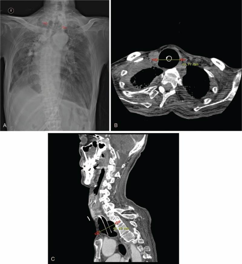
(A) Anteroposterior radiograph of the chest shows marked tracheomegaly (arrows). Computed tomography scans in axial (B) and sagittal (C) planes show marked tracheomegaly at the level of the tracheotomy tube cuff (arrows).

(A) Anteroposterior radiograph of the chest shows marked tracheomegaly (arrows). Computed tomography scans in axial (B) and sagittal (C) planes show marked tracheomegaly at the level of the tracheotomy tube cuff (arrows).