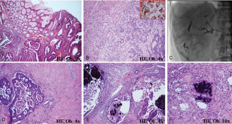
FIGURE 1.

In a patient with rectal adenocarcinoma (A) and liver metastases (B) marked by keratin 20 (B-frame), at 6 months after TACE (C), large hyalinized areas (D), and necrosis (E) can be seen in the hepatic metastasectomy specimen. The chemotherapic blue crystals can be observed inside the glandular lumen (E) and intravascular (F).
