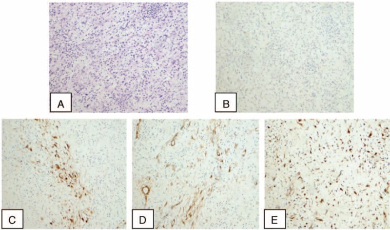
FIGURE 2.

A, The histopathologic examination demonstrates an abnormal proliferation of spindle cells in a fascicular and myxoid pattern with no significant nuclear atypia accompanied by inflammatory cells (hematoxylin and eosin stain, original magnification, ×100). Immunohistochemical appearance shows that calponin, anaplastic lymphoma kinase, smooth muscle actin, and vimentin are positive in inflammatory myofibroblastic tumor of this case (B-E ×100).
