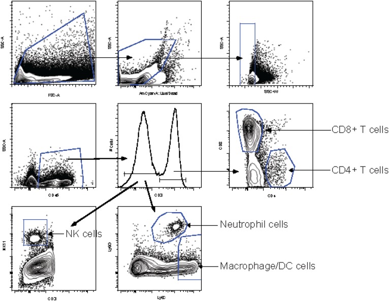
Fig. S4.
Gating strategy to identify immune cells in the brain and spleens. Single-cell suspensions were gated, excluding very small cells or debris, and cell doublets were excluded by side-scatter width. The LIVE/DEAD aqua dye was used to label dead cells, and only living cells were gated. The pan leukocyte marker CD45.2 was used to gate on leukocytes. Cells were gated on CD3+ cells that include CD4+ and CD8+ T cells, NK T cells, and γδ T cells. CD3+ cells were gated further on CD8+ and CD4+ cells. The CD3− gate was used to subset cells further into neutrophils (Ly6G+, Ly6C+), macrophages/dendritic cells (DC cells) (Ly6G−, Ly6C+), and NK cells (NK1.1+). Leukocytes per spleen and brain preparation were counted on a hemocytometer, and the numbers of each cell type were calculated.

