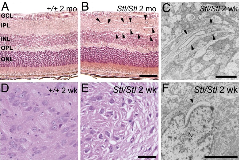
Fig. 3.
Aberrant membrane structures caused by the Stellar mutation. (A and B) Hematoxylin and eosin-stained retina from 2-mo-old wild-type (A, +/+) and homozygous mutant (B, Stl/Stl) mice. Retinal layers are labeled as follows: ganglion cell layer (GCL), inner plexiform layer (IPL), inner nuclear layer (INL), outer plexiform layer (OPL), and outer nuclear layer (ONL). (Scale bar, 50 µm.) (C) Electron micrograph of an area in the Stl/Stl retina. (Scale bar, 5 µm.) (D and E) H&E-stained sections showing the thalamus of 2-wk-old wild-type (D, +/+) and homozygous mutant (E, Stl/Stl) mice. (Scale bar, 50 µm.) (F) Electron micrographs of 2-wk-old homozygous mutant (Stl/Stl) mice showing abnormal membrane structures in the thalamus. Note the perinuclear membrane structures. N, nucleus. (Scale bar, 5 µm.) Abnormal membrane structures are marked by arrowheads (B, C, and F).

