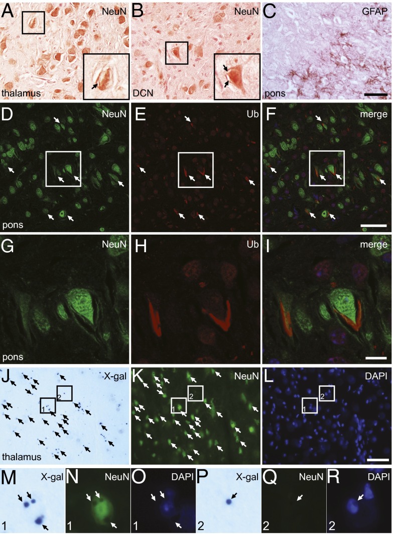
Fig. 5.
Neuronal localization of abnormal membrane structures in the Stellar brain. (A–C) Brain sections immunostained with antibodies against NeuN (A and B) and GFAP (C). Areas in thalamus (A), deep cerebellar nuclei (DCN, B), and pons (C) are shown. Areas enclosed by rectangles in A and B are also shown in detail in Insets. Note the intracellular abnormal membrane structures (arrows). (C) GFAP-stained brain sections showing an area in pons. (Scale bar, 50 µm.) (D–I) Most ubiquitin-positive structures in the Stellar brain are neuronal. Sections were incubated with antibodies against NeuN (D) and ubiquitin (E). An area in pons is shown. The merged image is shown in F. Ubiquitin-positive structures are marked (arrows). The enclosed area is also shown in detail (G–I). (Scale bar, 50 µm in D–F and 10 µm in G–I). (J–L) X-gal–positive cells in the Sptssb knockout mouse are mostly neurons. An area of the thalamus is shown. X-gal–stained cryosection (J) was also incubated with a NeuN antibody (K), which labels neurons, and counterstained with DAPI (L). Cells positive for both X-gal stain and NeuN are marked with arrows in J and K. Areas enclosed by rectangles are enlarged in M–R. Area 1 (M–O) shows a typical neuron positive for both X-gal (Sptssb, M) and NeuN (N). Area 2 (P–R) shows a rare case in which an X-gal–positive cell (P) is negative for NeuN (R). (Scale bar, 50 µm.)

