Fig. S2.
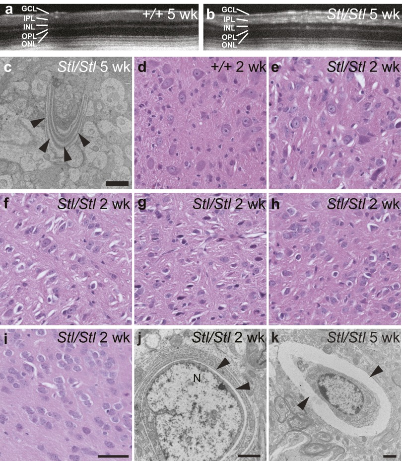
The Stellar mutation causes accumulation of abnormal membranous structures in the retina and the brain. (A and B) Optical coherence tomography (OCT) of 5-wk-old wild type, +/+ (A) and Stl/Stl (B) showing the light-reflecting flecks in the mutant eye. Retinal layers are labeled as in Fig. 3. (C) An abnormal membrane structure in 5-wk-old Stl/Stl retina shown by electron micrograph. (Scale bar, 2 µm.) (D–I) H&E staining of the deep cerebellar nuclei (D and E), pons (F), and medulla (G) in the brainstem, midbrain (H), and periform cortex (I) from 2-wk-old wild-type, +/+ (D) and homozygous Stl/Stl mutant (E–I) mice. (Scale bar, 50 µm.) (J and K) Electron micrographs of cells in the thalamus (J) and brainstem (K) from 2-wk-old (J) and 5-wk-old (K) homozygous Stl/Stl mutant mice, show cavities (marked by arrowheads) juxtaposed to the nucleus (J) or surrounding the cell (K). (Scale bar, 2 µm.)

