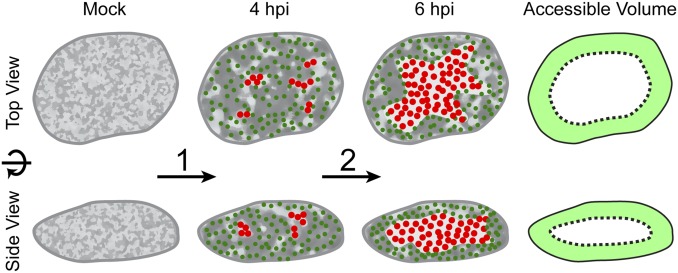
Fig. 8.
Viral remodeling of the nuclear architecture allows efficient flux of nuclear capsids in the absence of a directed transport system. Schematic depiction of the spatial changes during PRV infection that allow capsids to diffuse efficiently to site of nuclear egress. First (1), the interchromatin compartments enlarge, which leads to an increase in corral size (light gray regions). Capsids form and populate the enlarged corrals (green dots; 4 hpi). Over time (2), replication compartments form (red dots) enlarge and coalesce. At 6 hpi, chromatin is marginalized and most of the chromatin-depleted region in the center of the nucleus is occupied by the replication compartment. Capsids now mainly move in the peripheral corrals near the nuclear membranes, bringing them nearer to sites of egress. The accessible volume fraction is depicted as light green area, showing that a large fraction of the nuclear volume has access to nuclear membranes in 3D.

