Fig. 4.
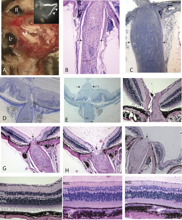
Ocular histopathology. (A) Gross dissection specimen of a 24-mo-old transgenic mouse with marked thinning of the entire left optic nerve (arrowheads) from the globe to the optic chiasm. (B and C) Longitudinal sections of atrophic optic nerve (arrows) (B) and the optic nerve (arrows) of an age-matched normal mouse (C). (D and E) Optic nerve head swelling 1 mo (D) and 3 mo (E) after birth. (F) A normal optic nerve head. (G–I) Loss of the optic nerve head tissues (arrow) at age 22 mo (G and H) and 26 mo (I). (J–L) Longitudinal retinal sections of a normal mouse (J) and transgenic mice at 22 mo (K) and 26 mo (L) of age. Arrows point to cells in the ganglion cell layer. (Scale bars, 100 µm.) INL, inner nuclear layer; ONL, outer nuclear layer.

