Fig. 1.
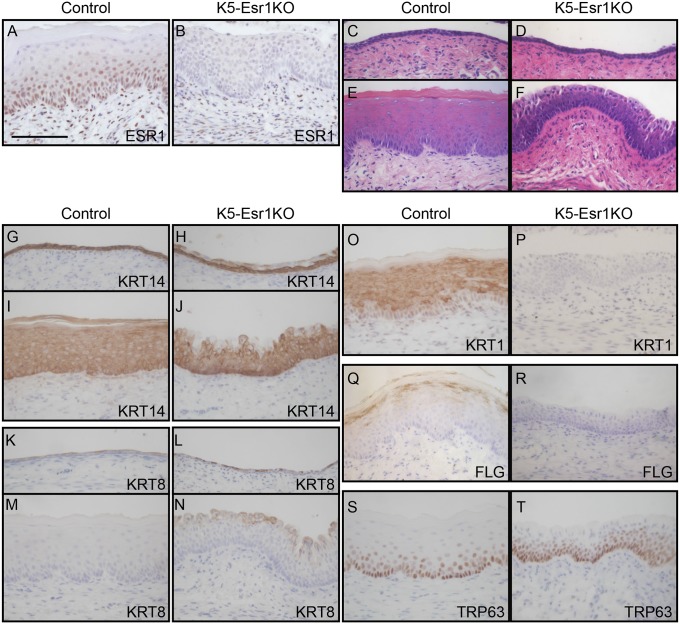
Effects of epithelial cell-specific ablation of Esr1 on the mouse vagina. ESR1 protein expression was evaluated by immunohistochemistry in control (A) and K5-Esr1KO mice treated with E2 (B), indicating successful deletion of Esr1 specifically in the epithelial cells in the K5-Esr1KO mice. The vaginal epithelium of 8-wk-old OVX control (C) and K5-Esr1 (D) mice. E2 treatment induces vaginal epithelial stratification and keratinized differentiation in the control (E), but fails to such differentiation in the vaginal epithelium of K5-Esr1KO mice (F). Expression pattern of cell differentiation markers in vaginal epithelium of OVX (G, H, K, and L) and E2-treated (I, J, M–T) mice. Immunohistochemical staining for KRT14 (G–J), KRT8 (K–N), KRT1 (O and P), FLG (Q and R), and TRP63 (S and T). In K5-Esr1KO mice, differentiating cell-specific markers KRT1 and FLG are not expressed, even after E2 treatment (C–F). (Scale bar, 100 µm.)

