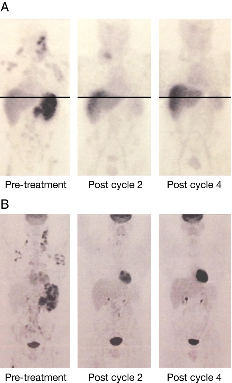
Fig. 2.
Clinical response of a patient with HL was demonstrated with 111In-daclizumab and FDG-PET. (A) 111In-daclizumab maximal intensity projection images (MIP) SPECT imaging studies of patient 1. (Left) At the time of the initial treatment with 90Y-daclizumab, there is localization in lymph nodes, bone, and spleen. (Center) After two treatments a decrease is seen in spleen, bone, and nodal uptake (the new uptake in the right base of the neck is related to central line placement). (Right) There is resolution of all abnormal uptake after four cycles. Findings with 111In-daclizumab were congruent with FDG findings. Note that the chest and abdomen/pelvis views were obtained in two separate acquisitions with slight overlap and then spliced together manually for display purposes. (B) Corresponding FDG-PET scans. (Left) Scan before treatment showing involvement in lymph nodes, spleen, and bone. (Center) At the time of the second treatment most of disease has resolved, with the exception of some nodes below the diaphragm. (Right) The images at the time of fourth treatment show complete resolution of disease with a progression-free survival of 400 d.

