


Figure 2.
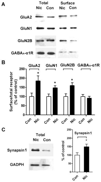
Extended nicotine exposure increases surface expression of glutamate receptor subunits on hippocampal neurons in culture. (A) Neurons were treated with control medium (Con) or with medium containing nicotine (Nic) for 7 days and then biotinylated, solubilized, and immunoblotted for GluA2, GluN1, GluN2B, or GABAA α1 receptors in aliquots of whole extract (left, total) or in the biotinylated fraction (right, surface). (B) Quantification of biotinylated surface components recovered on immunoblots from control (Con) and nicotine-treated (Nic) samples, normalized for the amount present in controls. GluA2, *p ≤ 0.02, n = 8; GluN1, *p ≤ 0.03, n = 5; GluN2B, *p ≤ 0.02, n = 5; GABAA α1, p ≥ 0.9, n = 5. (C) Synapsin 1 in whole extracts from control (Con) and nicotine-treated (Nic) cultures probed on immunoblots (GAPDH for reference), quantified, and normalized for control values (right; *p ≤ 0.02; n=5).