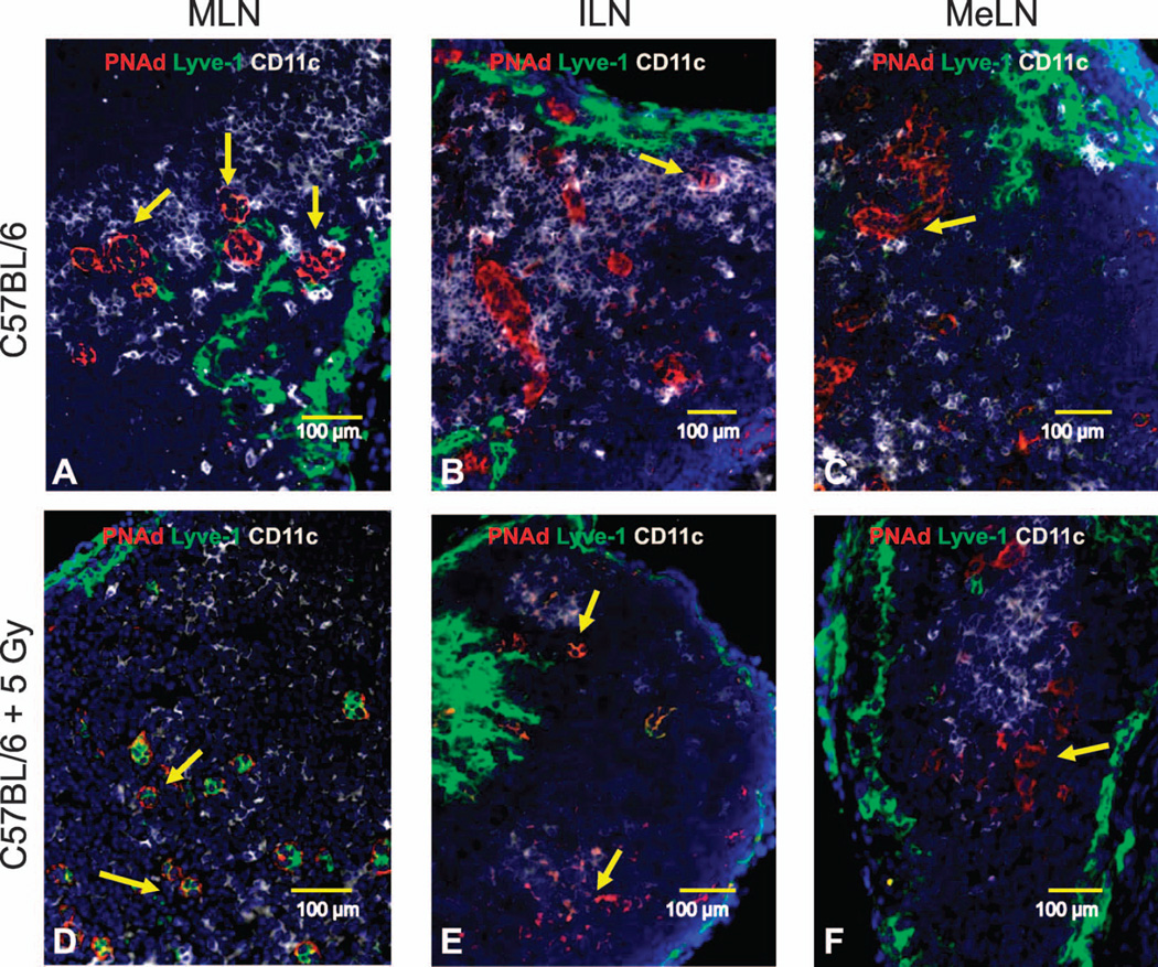
FIG. 5.
SLO structure was assessed in neonate mice 7 days postirradiation (frozen sections stained with fluorescent antibodies). Representative 200× pictures for PNAd+ HEV (red), Lyve-1+ lymphatics (green) and CD11c+ dendritic cells (white) in mediastinal lymph nodes [MLN; panel A (sham irradiated) and panel D (5 Gy TBI)], inguinal lymph nodes [ILN; panel B (sham irradiated) and panel E (5 Gy TBI)] and mesenteric lymph nodes [MeLN; panel C (sham irradiated) and panel F (5 Gy TBI)] are shown. Yellow arrows identify PNAd+ HEV.

