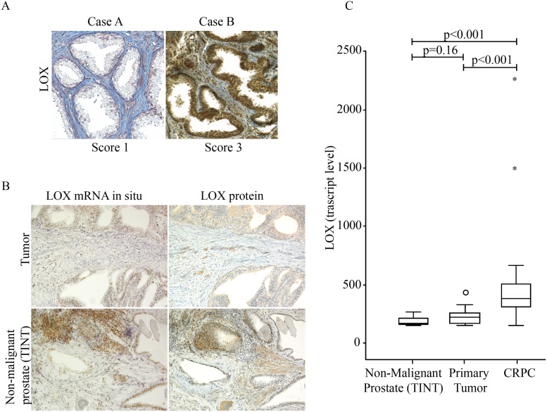
Fig 1. Lysyl Oxidase (LOX) expression in malignant and non-malignant human prostate tissue.
(A) Representative immunohistochemical staining of LOX (brown) in sections of non-malignant prostate tissue specimens (TINT epithelium and TINT stroma) from two prostate cancer patients (original magnifications x 200). Case A show weak epithelial staining (score 1) and Case B strong epithelial staining (score 3). (B) Consecutive sections from non-malignant prostate tissue stained with in situ hybridization or immunohistochemistry for LOX. Note that mRNA (brown dots) and protein (brown) expression in individual glands were related, in the stroma in contrast mRNA was generally low but protein often detected (original magnifications x200). (C) LOX mRNA expression in non-malignant prostate tissue (TINT) (n = 12), in primary prostate tumor tissue (n = 12), and in castration-resistant bone metastases (CRPC) (n = 30) using Illumina gene expression array.

