
FIG 3.
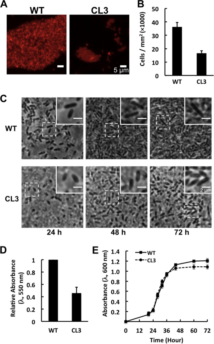
R. sphaeroides strain CL3 forms defective biofilms. (A) Representative fluorescence micrographs of R. sphaeroides wild-type (WT) and CL3 biofilms grown on a chamber slide with hydrophobic plastic surfaces for 72 h at 30°C in Sistrom's succinate medium containing 5 μg/ml Nile Red. (B and C) R. sphaeroides WT and CL3 cells were grown on a chamber slide with hydrophobic plastic surfaces at 30°C in Sistrom's succinate medium. Images were acquired at 1 (see Fig. S3 in the supplemental material), 24, 48, and 72 h after incubation by phase-contrast bright-field microscopy. Insets are magnified views of representative cells that demonstrate changes in cell length. Surface-attached cells were counted at 1 h. Shown are mean values ± standard deviations obtained from three independent experiments. (D) Quantification of R. sphaeroides WT and CL3 biofilms grown in wells of a polystyrene microtiter plate for 72 h at 30°C in Sistrom's succinate medium and then labeled with CV. Biofilm formation was quantified by measuring the absorbance of CV at a wavelength of 550 nm. Shown are mean values ± standard deviations obtained from three independent experiments, each performed in eight replicates. (E) Growth curves of R. sphaeroides WT and CL3 cells grown with shaking in glass test tubes at 30°C in Sistrom's succinate medium. Shown are mean values ± standard deviations obtained from three independent experiments.