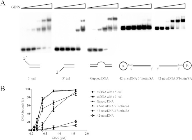
FIG 2.
Binding of SsoGINS to DNAs of different structures. SsoGINS was mixed with a 32P-labeled DNA fragment containing a 42-bp dsDNA region with a 5′ tail (D2) or a 3′ tail (D3) (see Table S1 in the supplemental material), a 32P-labeled partial dsDNA fragment containing a 20-dT region in the middle (D6) (see Table S1), a 42-nt ssDNA with 5′ biotin-SA, or a 42-nt ssDNA with 3′ biotin-SA. The protein-DNA complexes were subjected to polyacrylamide gel electrophoresis. The gel was exposed to X-ray film (A) and quantified by phosphorimaging (B). Concentrations of SsoGINS were 0, 0.1, 0.2, 0.4, 0.8, and 1.6 μM, indicated by the triangles above the lanes. Data shown in panel B represent an average of three independent measurements.

