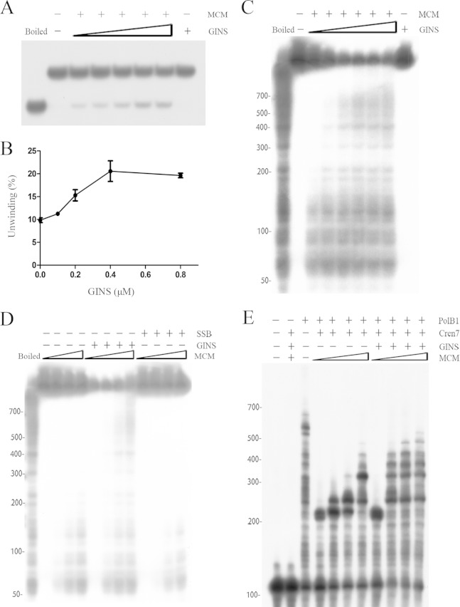
FIG 6.
Processivity of SsoMCM. (A and B) DNA unwinding by SsoMCM. SsoMCM (0.2 μM) was incubated with a radiolabeled oligonucleotide substrate (D4) (see Table S1 in the supplemental material) in the presence of increasing amounts of SsoGINS. Reaction products were subjected to electrophoresis in a polyacrylamide gel in 1× TBE buffer. The gel was exposed to X-ray film (A) and quantitated by phosphorimaging (B). Data shown in panel B represent an average of three independent measurements. Left lane, boiled substrate; second lane from left, no protein added; far-right lane, 0.8 μM SsoGINS. Concentrations of SsoGINS were 0, 0.1, 0.2, 0.4, and 0.8 μM, indicated by the triangles above the lanes. (C) Effect of SsoGINS on the processivity of SsoMCM. SsoMCM (0.2 μM) was incubated with a radiolabeled M13-based substrate in the presence of increasing amounts of SsoGINS. Reaction products were subjected to electrophoresis in 5% polyacrylamide gel in 1× TBE buffer. Left lane, boiled substrate; second lane from left, no protein added; far-right lane, 1.2 μM SsoGINS. Concentrations of SsoGINS were 0, 0.1, 0.2, 0.4, 0.8, and 1.2 μM, indicated by the triangles above the lanes. (D) Effect of SsoSSB on the processivity of SsoMCM. Various amounts of SsoMCM were incubated with the radiolabeled M13-based substrate in the presence of SsoGINS (1 μM) or SsoSSB (1.6 μM). Reaction products were subjected to electrophoresis in 5% polyacrylamide gel in 1× TBE buffer. The gel was exposed to X-ray film. Left lane, boiled substrate. Concentrations of SsoMCM were 0, 0.1, 0.2, and 0.4 μM, indicated by the triangles above the lanes, for each set of conditions. (E) Coupled DNA helicase and polymerase assays. SsoMCM was preincubated for 5 min with a 32P-labeled oligonucleotide (S13) (see Table S1) annealed to a 200-nt circular ssDNA in the presence or absence of SsoGINS (1 μM). Cren7 (1 μM) was added, and the incubation was continued for an additional 5 min. SsoPolB1 (25 nM) was then added. After further incubation, reaction products were processed for electrophoresis in polyacrylamide gel containing 7 M urea in 1× TBE buffer. The gel was exposed to X-ray film. Concentrations of SsoMCM were 0, 0.1, 0.2, and 0.4 μM, respectively, as indicated by the triangles above the lanes, for the conditions with SsoGINS and without SsoGINS. Values at the left of panels C to E are nucleotides.

