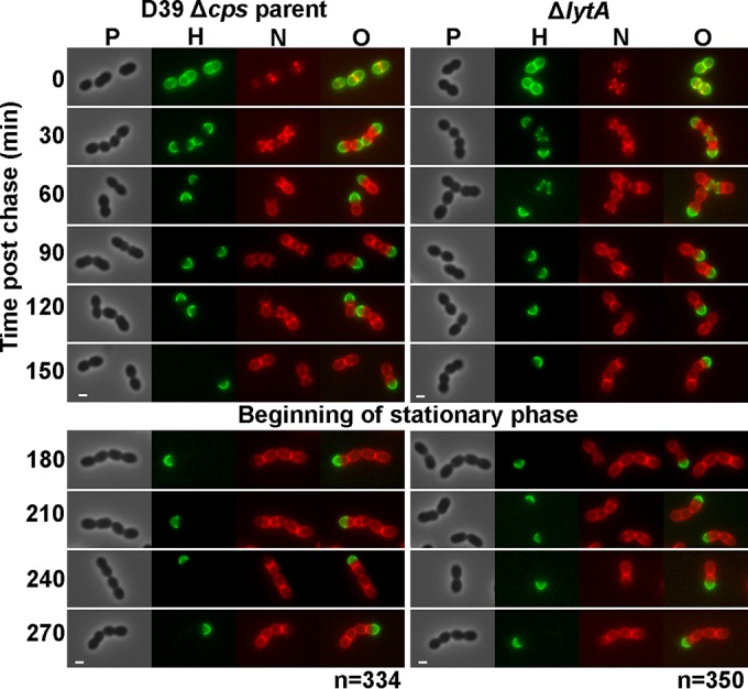
FIG 2.

Persistence of hemispheres of stable old PG in exponentially growing and early-stationary-phase cells of S. pneumoniae detected by FDAA long-pulse–chase–new-labeling experiments. Parent strain IU1945 (D39 Δcps) and an isogenic lytA mutant (IU3900) were grown exponentially in BHI broth, labeled with HADA (pseudocolored green; old PG) for about 3 generations, washed, and then chased in the presence of a second color of FDAA, NADA (pseudocolored red; new PG synthesis) as described in Materials and Methods. Live cells were imaged by epifluorescence phase-contrast microscopy at the indicated time points after the removal of HADA (chase) and the addition of NADA. Minimal variation in labeling patterns was observed for >330 individual cells in diplococci or short chains of each strain examined in microscopic fields at different time points after the start of the chase/NADA labeling, and representative images are shown. Labeling of the parent strain was done numerous independent times, and labeling of the lytA mutant was done twice with similar results. Scale bar = 1 μm. P, phase-contrast image; H, HADA labeling (old PG); N, NADA labeling (new PG synthesis); O, overlay of H and N images. Quantitation indicating hemispherical labeling is in the text.
