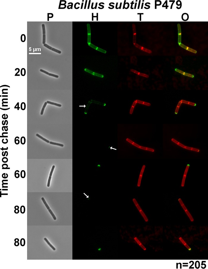
FIG 4.
Rapid turnover of sidewall PG in exponentially growing cells of B. subtilis detected by FDAA long-pulse–chase–new-labeling experiments. B. subtilis wild-type laboratory strain P479 was grown exponentially in LB broth (±1% [wt/vol] glucose; results without glucose are shown), labeled with HADA (pseudocolored green; old PG) for about 3 generations, washed, and then chased in the presence of a second color of FDAA, TADA (pseudocolored red; new PG synthesis) as described in Materials and Methods. Live cells were imaged by epifluorescence phase-contrast microscopy at the indicated time points after the removal of HADA (chase) and the addition of TADA. Minimal variation in labeling patterns was observed for ≈200 individual cells examined in microscopic fields at different time points after the start of the chase/TADA labeling, and representative images are shown. Labeling was done twice (once with added glucose and once without) with similar results. Scale bar = 5 μm. P, phase-contrast image; H, HADA labeling (old PG); T, TADA labeling (new PG synthesis); O, overlay of H and T images. The arrow at 40 min of chase indicates faint residual old (green) sidewall PG remaining. Arrows at 60 and 80 min indicate faint inert PG that likely resulted from partial synthesis. See the text for additional details.

