FIG 5.

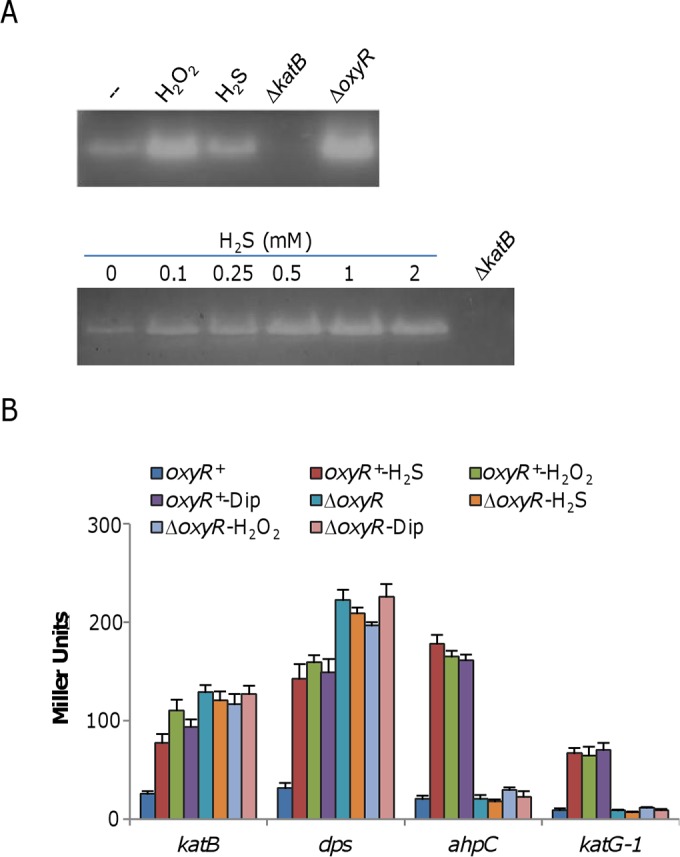
H2S induces the OxyR-mediated stress response. (A) CAT staining analysis. Cells were harvested just prior to (−) and 30 min after the addition of 0.25 mM H2S or 0.1 mM H2O2 (top). Protein samples of ∼10 μg from the cell lysates indicated were separated by native PAGE and stained for CAT activity. The ΔkatB and ΔoxyR mutant strains (constitutive high-level expression) were used as negative and positive controls. In the analysis shown at the bottom, various concentrations of H2S were examined for the ability to induce expression of the katB gene. (B) Impact of H2S on the expression of four members of the OxyR regulon. β-Galactosidase assays were carried out with lacZ reporter vectors. Cells grown to mid-log phase were treated with the chemicals indicated for 30 min and then harvested for the assays. Concentrations: H2S, 0.25 mM; H2O2, 0.2 mM; dipyridyl (Dip), 0.25 mM. β-Galactosidase activities are reported as the mean ± SD (n = 4). Similar results were obtained by qRT-PCR assay (see Fig. S3 in the supplemental material).
