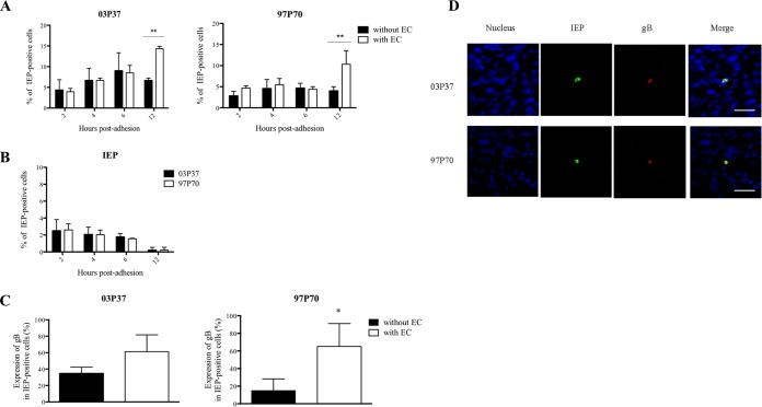
FIG 4.
Expression kinetics of EHV-1 proteins in blood CD172a+ cells upon adhesion to EC. (A) Expression kinetics of EHV-1 IEP in adherent blood CD172a+ cells. Cells were inoculated with EHV-1 strain 03P37 or 97P70 (MOI = 1) and cocultured with immortalized EC monolayers for 2, 4, 6, and 12 h. As a control, EHV-1-inoculated CD172a+ cells were incubated on plastic (without EC). (B) Expression kinetics of EHV-1 IEP in the nonadherent CD172a+ cell fraction after 2, 4, 6, and 12 h coculture with immortalized EC. (C) Expression of EHV-1 gB protein in infected CD172a+ cells after 12 h coculture with immortalized EC in the presence of neutralizing EHV-1 antibodies. Three independent experiments were performed, and the data are represented as means plus SD. A two-way ANOVA test was performed to evaluate significant differences from the control (*, P < 0.05; **, P < 0.01). (D) Double immunofluorescence of EHV-1 IEP (green) and gB (red) proteins in blood CD172a+ cells adherent to immortalized EC for 12 h in the presence of neutralizing EHV-1 antibodies. Nuclei were counterstained with Hoechst (blue). All the confocal images represent merged z-stacks. Scale bars = 50 μm.

