FIG 1.
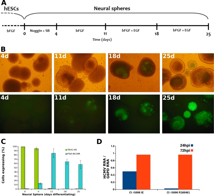
Transition toward HCMV susceptibility in early hESC-derived NPC. (A) Schematic presentation of induction of hESC differentiation into NPC. Undifferentiated hESC were maintained in culture medium supplemented with basic fibroblast growth factor 2 (bFGF). Upon initiation of neural differentiation (days 0 to 4), bFGF was withdrawn from the culture medium. The medium was supplemented during days 0 to 4 with two inhibitors of the SMAD signaling pathway (Noggin and SB-431542). From day 5, the SMAD signaling inhibitors were removed, and the medium was supplemented with bFGF. From day 12, the medium was further supplemented with epidermal growth factor (EGF) (25, 28). (B) Neurospheres formed at the indicated time points post-induction of differentiation were infected with HCMV strain TB40/E expressing the late gene pp150 fused to GFP (multiplicity of infection of 1 PFU/cell) and visualized at 7 days postinfection by inverted-phase (top row) and fluorescence (bottom row) microscopy. Similar findings were obtained using HCMV strain TB40/E expressing immediate-early GFP (not shown). (C) Expression of cell differentiation markers by neurospheres formed at the indicated times post-induction of differentiation, as analyzed by immunostaining and flow cytometry. (D) Neurospheres at day 11 post-induction of differentiation were infected with the low-passage-number clinical strain CI5006, recovered from the urine of a congenitally infected newborn. Shown are the viral IE-1 and late RNA R160461 mRNA levels, determined as described previously (27) at the indicated times postinfection and normalized by the housekeeping gene for glucose-6-phosphate dehydrogenase (G6PD) (similar results were obtained with three additional low-passage-number clinical isolates).

