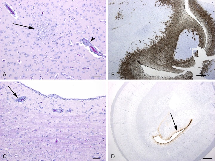
FIG 5.
Comparison of histopathology and immunohistochemistry findings in brains of P6 rH5N3 (A and B)- and P0 rH5N3 (C and D)-inoculated birds at 3 dpi. (A) Perivascular cuffing with mononuclear inflammatory cells (arrowhead) was observed throughout the brain. Note area of gliosis and necrosis (arrow). Bar, 50 μm; H&E stain. (B) Extensive positive immunostaining for influenza A virus antigen. Bar, 500 μm. (C) Perivascular cuffing was observed only in the periventricular area of one bird (arrow). Bar, 50 μm; H&E stain. (D) Detection of viral antigen was limited to the ependymal cells lining the ventricles. Bar, 500 μm.

