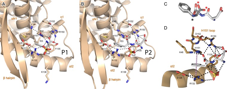
FIG 2.
Details of the P-CTD binding pocket and interaction network with the N-NTD. (A and B) Enlarged views of the binding of phenylalanine (P1) (A) and Asp-Phe (P2) (B), corresponding to the P C terminus. The N-NTD is in tan and shown in cartoon representation, with secondary structures labeled. Residues involved in binding through electrostatic (dashed lines) or van der Waals interactions are shown in stick representation and colored by atom type, with carbons in tan. P1 and P2 are in stick representation, with the same color scheme as in Fig. 1. (C) Superposition of P2 and P1 conformations. Atoms with conserved polar contacts are indicated by asterisks, notably, two oxygen atoms (red asterisks). (D) Details of the double stacking interaction between the aromatic ring of P2 and the side chains of H151 and R132, with electrostatic interactions between P2 and the N-NTD indicated by black dashed lines. The normal axis to the P-F24 ring plane is indicated by the violet dotted line.

