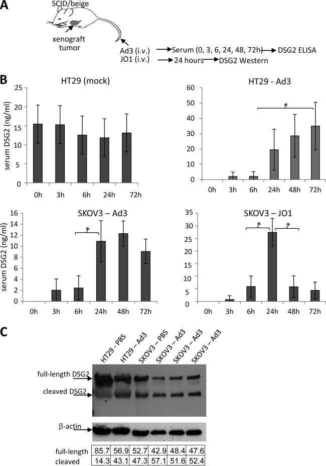
FIG 3.
DSG2 shedding in vivo in mice with tumors derived from human cancer cell lines. (A) Experimental design. Immunodeficient CB17 SCID/beige mice with established subcutaneous human xenograft tumors (∼500 mm3) were injected intravenously with Ad3 virus (2 × 10⁹ PFU per mouse) or JO1 (2 mg/kg). Preinjection serum samples (0 h) and samples harvested at 3, 6, 24, 48, and 72 h after Ad3 injection were analyzed by ELISA for DSG2 ECD concentrations. (B) Serum DSG2 levels in mice with xenograft tumors derived from SKOV3 or HT29 cells. The left upper panel shows background DSG2 serum levels in mice with HT29 tumors. In all other panels, background (preinjection) DSG2 levels were subtracted from the levels measured after Ad3 or JO1 injection (n = 5). *, P < 0.01. (C) DSG2 Western blot of tumor lysates after Ad3 injection. Mice with HT29- or SKOV3-derived xenograft tumors were injected with Ad3, and tumors were harvested 24 h later. 6D8 antibody was used as a probe. DSG2 band signals were normalized to β-actin signals and are expressed as a percentage of combined (DSG2) intensities (according to the table below the blot). Each lane represents an individual tumor.

