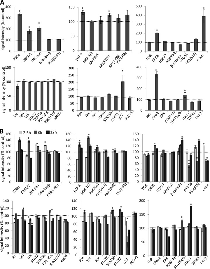
FIG 4.
Analysis of phosphokinase activation after incubation of A549 cells with Ad3 and JO1. (A) A549 cells were incubated with Ad3 at an MOI of 100 PFU/cell for 1 h on ice to allow for attachment. Virus was then removed, and cells were incubated for 2.5 h at 37°C. Cell lysates were subjected to hybridization on filters containing antibodies that are able to detect relative levels of phosphorylation of 43 different kinase phosphorylation sites (Proteome Profiler antibody array; R&D Systems). Signals were quantitated and plotted, taking the signals from mock-infected cells as 100%. *, P < 0.01 (n = 3 independent experiments). (B) A549 cells were incubated with 1 μg/ml of JO1 and harvested at 2.5, 8, and 24 h later. As a control, A549 cells were incubated with Ad35 fiber knob (1 μg/ml) and harvested at 2.5 h. JO1 signals were normalized to Ad35 fiber knob signals (taken as 100%) (n = 3). eNOS, endothelial nitric oxide synthase.

