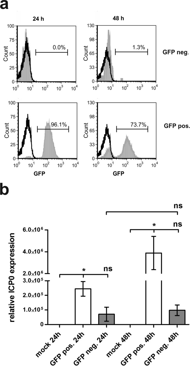
FIG 3.
GFP-negative bystander DCs are not secondarily infected. Mature DCs were either mock or HSV-1 infected. At 16 h postinfection, cells were sorted into GFP+ and GFP− cells and analyzed directly or after an additional 24 h of incubation. (a) Cells were analyzed for their GFP expression using flow cytometry. Mock controls are depicted as black histograms; GFP− (upper panel) and GFP+ (lower panel) cells are depicted as filled light gray histograms. The percentage of GFP+ cells is depicted in each histogram. (b) qRT-PCR analyses were performed to assess relative ICP0 expression levels. S14 and GAPDH served as controls. The experiment was carried out three times with cells from different healthy donors. Significant changes (P < 0.05) are indicated by asterisks. Nonsignificant changes (P > 0.05) are depicted as “ns.” neg., negative; pos., positive.

