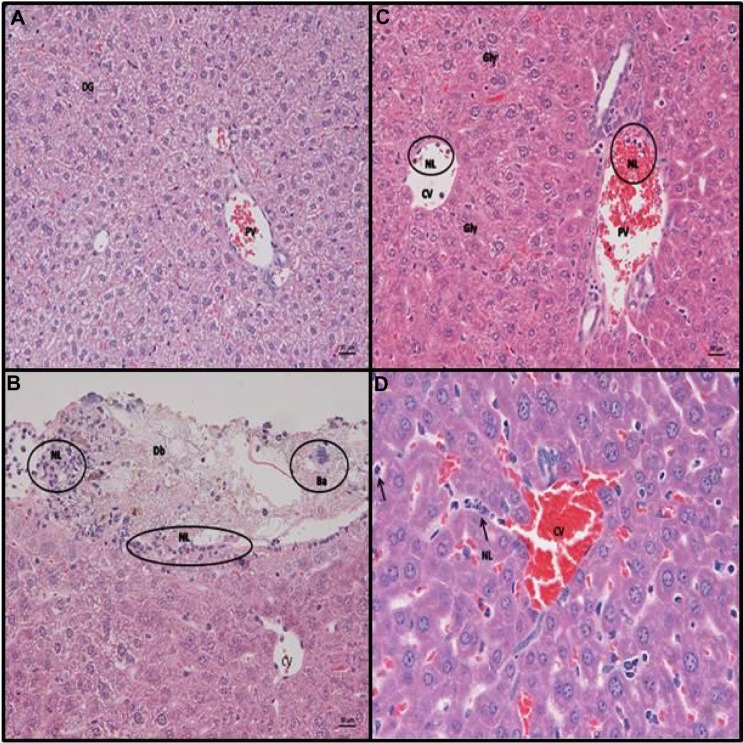
Figure 4.
Representative slides from liver for animals receiving IP saline/lPs/stool suspension.
Notes: Hematoxylin and eosin staining of C57BL/6 mouse liver at 6 hours post (A) intraperitoneal (IP) injection with saline. Normal histology noted, DG (B) IP inoculation of stool (5 µl/g body weight). Liver parenchyma with capsule, exhibiting focal serosal attachment of nls and cellular debris (perihepatitis). Underlying the infiltrate is a thin layer of subserosal neutrophils. Ba from the stool suspension can also be seen. (C and D) IP injection with LPS (25 mg/kg). Liver parenchyma with neutrophils in the lumen of CVs and PVs. Closer view of the liver parenchyma highlights the increased amount of neutrophils between hepatic cords. Hepatocytes do not exhibit cytoplasmic vacuolation, consistent with a diffuse loss of glycogen (scale bar =20 µm, A–C; scale bar =10 µm, D). Representative examples of neutrophils are circled “NL” in B and C and arrowed “NL” rolling along the hepatic chords in D. Bacterial colonies are circled “Ba” in B.
Abbreviations: Db, bacterial debris; Gly, glycogen; LPS, lipopolysaccharide; NLs, neurtrophils; CVs, central veins; PVs, portal veins; Ba, bacterial colonies; DG, diffuse glycogen.

