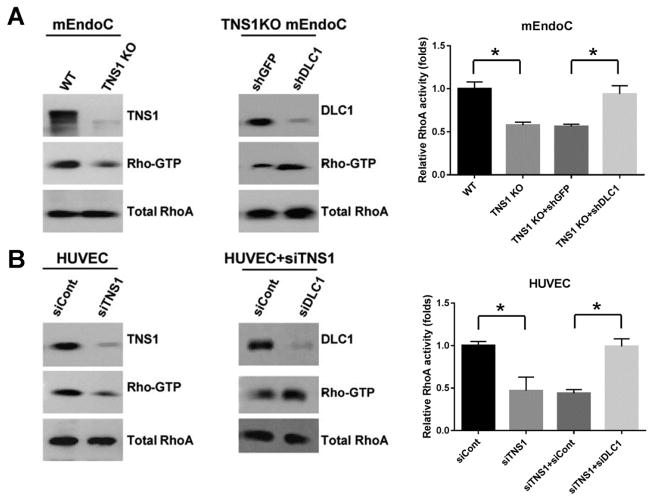
Figure 4. Lack of TNS1 expression reduces the level of active RhoA, which could be restored by further silencing of DLC1 in mouse endothelial cells.
(A) Primary endothelial cells (mEndoCs) isolated from WT or TNS1 KO mice were lysed for RhoA activity assays (left panel). TNS1 KO cells infected with either shGFP or shDLC1 lentivirus were analyzed for RhoA activities (right panel). (B) HUVEC transfected with control (siCont), TNS1 siRNA (siTNS1), or siTNS1 together with siCont or siDLC1 were lysed to determine RhoA activities. Whole cell lysates were immunoblotted with TNS1, DLC1, and RhoA antibodies to assess TNS1 or DLC1 depletion in cells and total RhoA levels were served as loading controls. Bar graph shows quantification of Rho-GTP. Shown are means ± SD for a minimum of three repetitions per transfection condition. P value was calculated by one-way ANOVA with Tukey-Kramer post hoc test. *, P < 0.01

