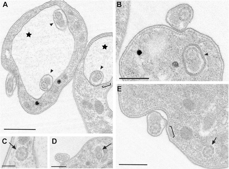
FIG 3.
Ultrastructural phenotype of TbMORN1-depleted cells. Shown are common morphological characteristics of TbMORN1-depleted cells (14-h point). Images were taken from 60-nm-thick resin sections contrasted with uranyl acetate and lead citrate. (A) Enlarged vacuoles (stars) and multiple axonemes with PFRs inside the pocket (arrowheads). The microtubule quartet seems unaffected (bracket; also in panel E). (B) Axoneme with PFR inside the flagellar neck region (arrowhead). (C to E) Intracellular axonemes with and without PFR (arrows). Scale bars: 1 μm (A and D), 500 nm (B and E), and 200 nm (C).

