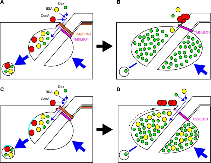
FIG 8.
Summary of observations and two possible interpretations. Shown are schematic representations of the posterior end of a trypanosome cell in longitudinal cross-section. The FPC is depicted in magenta (TbBILBO1). The TbMORN1 macromolecular complex is shown as an orange fishhook above the FPC. Dextran, BSA, and ConA are depicted, respectively, as green, yellow, red circles. Blue arrows indicate movement. Endocytosis and secretion are represented by large blue arrows. (A) Model 1. In uninduced cells, dextran, BSA, and ConA enter the FP (small blue arrows) and are taken up by endocytosis and trafficked to the endosomal-lysosomal system in transport vesicles. The secretory pathway returns membrane to the FP. (B) Following TbMORN1-directed RNAi, the FPC is intact but TbMORN1 is largely absent. BSA (fluid phase) and ConA (membrane bound) can no longer efficiently access the FP, and ConA accumulates close to the point of flagellum entry. Dextran can still access the FP and accumulates inside it. The rate of endocytosis is reduced (small blue arrow), while secretion is unaffected. (C) Model 2. The situation is the same as that depicted in model 1, except for the behavior of ConA. Here, ConA binds exclusively to glycoproteins present either within or adjacent to the FP in a defined patch (cyan, here shown inside the FP). (D) Following depletion of TbMORN1, the maintenance of subdomains is lost and the ConA-binding glycoprotein patch is displaced outside the FP (dashed arrow), where it accumulates ConA. The rate of endocytosis is reduced as in model 1, leading to an enlargement of the FP and an accumulation of both dextran and BSA within it.

