FIG 5.

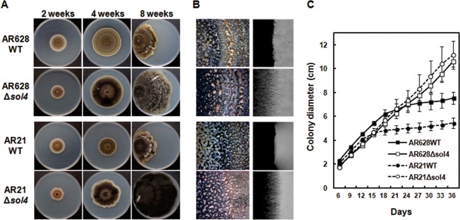
Colony morphology of WT and Δsol4 mutant strains. (A) Colony morphology of AR628 and AR21 WT strains and their corresponding Δsol4 mutants at 2, 4, and 8 weeks after incubation on PDA. For 8-week-old cultures, strains were grown from the left-hand edge of the plates. Note that the restricted colony size for the WT strains is due to the production and accumulation of solanapyrones. (B) (Left) Normal sporulation of both the WT strains and Δsol4 mutants at 3 weeks after incubation on PDA. The mass of spores oozing out from the pycnidia was visible on the colony surface of both strains. (Right) Inhibition of mycelial growth and the toxic effect of solanapyrone on mycelial growth are visible at the colony margins in the wild-type strain but not in the Δsol4 mutant strains. See panel A for the strains shown in the four panels in each column. (C) Growth curves of the WT strains and their corresponding Δsol4 mutants on PDA. Colony diameters were measured at 3-day intervals. Error bars are standard deviations (n = 5). The initial growth rates were similar among the WT and Δsol4 mutant strains in the first 2 weeks, but the growth rate became lower in the WT strain due to inhibition by solanapyrones.
