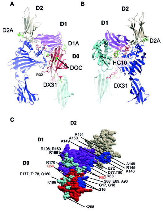
Figure 6. Model of KIR3DL2 binding to B27 free heavy chain dimers.

A Front and B reverse view of KIR3DL2 bound to B27 dimer. B27 heavy chains are shown in light and dark blue. The D0, D1 and D2 domains of KIR3DL2 are coloured red, purple and coral. The locations of HC10, DOC, D1A, D2A and DX31 antibody epitopes are indicated in green. R32, implicated in KIR3DL2 ligand binding is also indicated. C. The B27 footprint mapped to the surface of KIR3DL2 with residues coloured cyan or blue to indicate the respective heavy chain contacts. Residues incorporated in the heavy chain HC10 MAb epitope are in red type.
