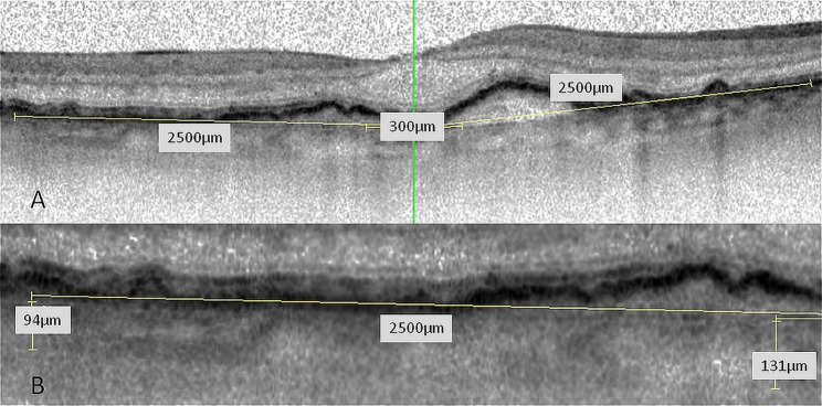
Figure 1.

(A) Optical coherence tomography (OCT) scan through the fovea; five locations (subfoveal, 300 µm and 2500 µm nasal and temporal to the fovea) were measured. (B) CT-measurements were performed by two independent readers. The imaging software of Spectralis OCT includes contrast enhancement, copying of overlays, side by side comparison of different OCT scans and flickering of two scans allowing for precise measurements.
